 02-056-3333


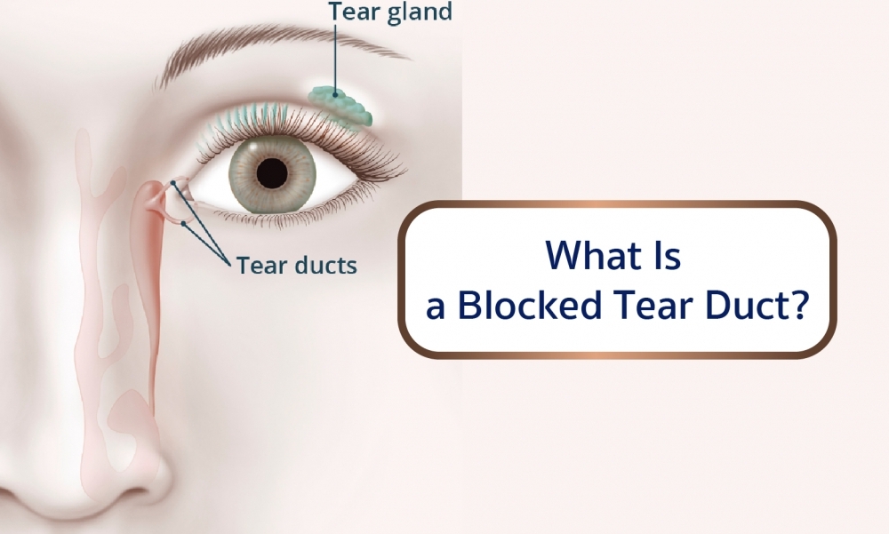
What Is a Blocked Tear Duct?
A blocked tear duct is when the eye’s drainage system for tears is either partially or completely obstructed. Tears cannot drain normally, causing a watery, irritated or chronically infected eye.
Most of your tears come from your lacrimal glands, which are located above each eye. The tears flow down the surface of your eye to lubricate and protect it, and then drain into tiny holes (puncta) in the corners of your upper and lower eyelids. The tears then travel through the small canals in the lids (canaliculi) to a sac where the lids are attached to the side of the nose (lacrimal sac), then down a duct (the nasolacrimal duct) before emptying into your nose, where they evaporate or are reabsorbed.
A baby can be born with a blocked tear duct (a congenital blocked tear duct). It is estimated nearly 20 percent of newborns have a blocked tear duct, but the condition usually resolves on its own within four to six months. In adults, the tear duct obstruction can result from an eye infection, swelling, injury or a tumor.
Blocked Tear Duct Causes
A blockage can occur at any point in the tear drainage system. When that happens, your tears don't drain properly, giving you watery eyes and increasing your risk of eye infections and inflammation.
Babies in utero have a thin membrane that seals the nasolacrimal duct. In newborns , a blocked tear duct may be the result of that membrane not opening as it should at birth.
Another cause of blocked tear duct may be chronic nose infections. Chronic sinusitis may irritate the tissues and form scars, which block the tear duct system.
Other causes of blocked tear duct:
Abnormal development of the skull and face (craniofacial abnormalities), like those in Down syndrome or other disorders, increases the likelihood of blockage of the tear ducts.
Age-related changes in older adults can cause blocked tear ducts, including narrowing of the punctal openings.
Nose trauma , such as a broken nose; scar tissue can block the tear duct.
Nose polyps , a growth from the lining of the nose (affecting some people who have nasal allergies), can obstruct the tear duct system
Conjunctivitis, infection and inflammation of the conjunctiva, the thin membrane covering the eye. In rare cases, the tear duct system may become infected and blocked, especially after some viral infections
Tumor , which may press on the tear duct system and prevent drainage.
If your eye has been watery and leaking or is continually irritated or infected, you should see your ophthalmologist.
Blocked Tear Duct Treatment
Sometimes, more than one treatment or procedure is needed before a blocked tear duct is fully opened. If an infection is suspected, your doctor will likely prescribe antibiotics.
Many babies with congenital blocked tear duct improve on their own in the first several months of life, after the drainage system matures or the extra membrane involving the nasolacrimal duct opens up. In some cases, your ophthalmologist may recommend that you use a special massage technique to help open up the membrane covering the lower opening into your baby's nose. He or she will demonstrate how to correctly do this massage.
The purpose of massage is to put pressure on the lacrimal sac to pop open the membrane at the bottom of the tear duct. This is most easily accomplished by placing your hands on each side of the baby’s face with your index finger(s) between the inner corner of the eye and the side of the nose, pressing in and down over the lacrimal sac for a few seconds. The massage should be done once in the morning and once in the evening, and each massage should be ten strokes each. It is best to do the massage during a diaper change.
In most cases of blocked tear ducts after a facial injury, the drainage system starts working again on its own a few months after the injury, and no additional treatment is necessary. Your ophthalmologist may recommend waiting a few months after the injury before considering surgery to open the blocked tear duct.
For infants and toddlers whose blocked tear ducts aren't opening on their own, or for adults who have a partially blocked duct or a partial narrowing of the puncta, a technique using dilation, probing and irrigation may be used. An instrument is used to enlarge (dilate) the punctal openings and a narrow probe is guided through the puncta, into the tear drainage system, then through the nasal opening and removed. The tear drainage system is flushed with a saline solution to clear out any residual blockage.
A balloon catheter dilation procedure opens tear drainage passages that are narrowed or blocked by scarring or inflammation. General anesthesia is used. A narrow catheter (tube) with a deflated balloon on the tip is guided through the lower nasolacrimal duct. The doctor then uses a pump to inflate and deflate the balloon along the drainage system.
With a procedure called stenting or intubation , tiny tubes are used to open up blockages and narrowing within the tear drainage system. Again, general anesthesia is usually used. Your ophthalmologist threads a very thin tube through one or both puncta in the corner of your eye, all the way through the tear drainage system and out through your nose. A tiny loop of tubing remains at the corner of your eye, but while it is visible, it's usually not bothersome. These tubes are generally left in for three to four months, and then removed.
Surgery is usually the preferred option for people who develop blocked tear ducts. It is also effective in babies and toddlers with congenital blocked tear ducts, though usually an option only after other treatments have been tried.
Dacryocystorhinostomy is the surgical procedure usually used to treat most cases of blocked tear ducts in adults and rarely in children. This technique creates a new route for tears to drain out through your nose normally again by developing a new connection between your lacrimal sac and your nose. This new route bypasses the duct that empties into your nose (nasolacrimal duct), which is typically the blockage site. Stents or intubation typically are placed in the new route while it heals, and then removed three or four months after surgery. The steps in this procedure will vary depending on your particular tear duct blockage.
Depending on the type of blockage, your surgeon may recommend creating an entirely new route from the inside corner of your eyes (puncta) to your nose, bypassing the tear drainage system altogether. This reconstruction of your entire tear drainage system is called conjunctivodacryocystorhinostomy .
To prevent postoperative infection and inflammation, you will need to use a nasal decongestant spray and eye drops. After about three to six months, your ophthalmologist will remove any stents that were put in place to keep the new channel open while healing.
If a tumor is causing your blocked tear duct, surgery may be performed to remove the tumor, or other treatments may be used to shrink it.
Information from American Academy of Ophthalmology
